Treatment
Retinal surgery repairs structural damage at the back of the eye, such as a retinal detachment, a macular hole, or scar tissue on the retina, when laser or injections alone are not enough. A detached retina is urgent and needs prompt assessment to give the eye the best chance of recovery.
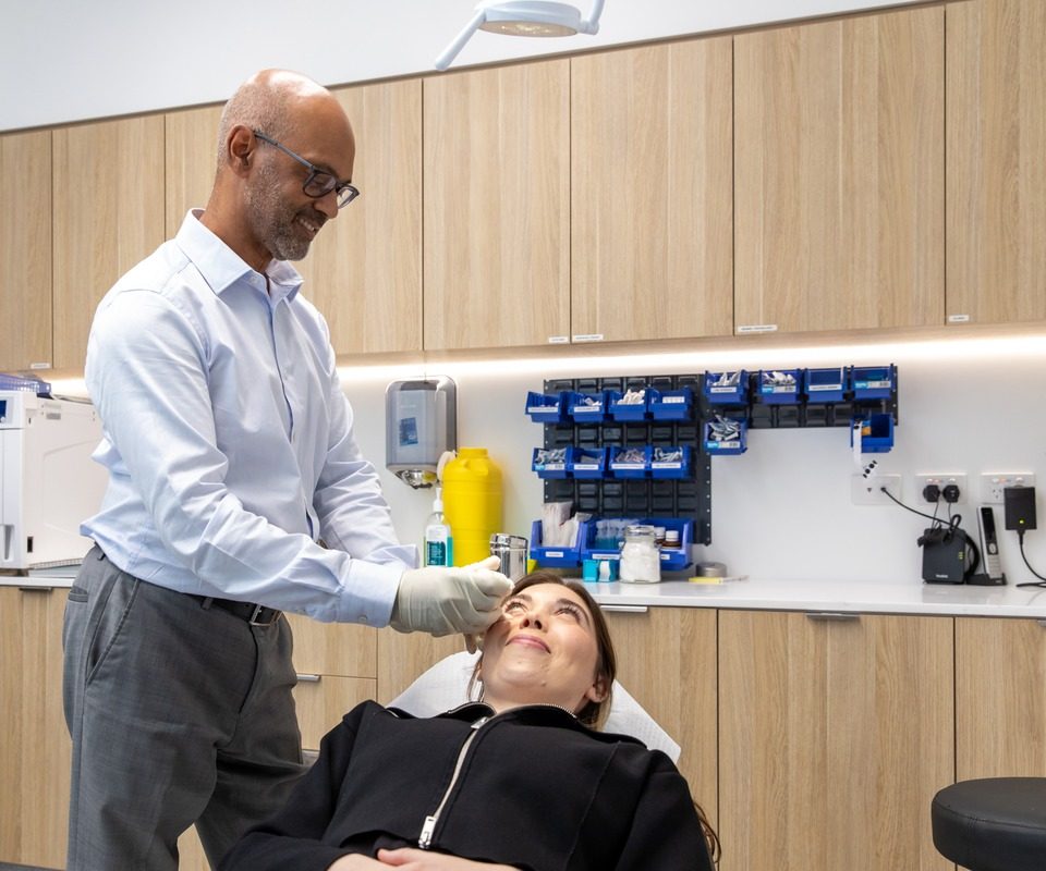
Retinal procedures are performed by our retina team at the facility selected for your operation. Epping Surgery Centre is our main day-surgery venue, while Sydney Eye Hospital, Macquarie University Hospital and Chatswood Private Hospital may also be used depending on the surgeon and procedure. Local or general anaesthesia may be used. Outcomes depend on the timing and extent of the problem.
Keyhole technique means the work is done through openings smaller than a pinhead, using very fine instruments
Gas or oil support is sometimes placed inside the eye to hold the retina while it heals, which can require a period of face-down positioning
No flying with a gas bubble is a firm rule until your surgeon confirms it has cleared, because cabin pressure changes can harm the eye
Same-day discharge is usual, with most people going home a few hours after the operation
Follow-up over weeks to months, since vision often keeps improving well after the eye itself has healed
At Eastwood Eye Surgery, our retina team assesses each eye individually. We discuss the most appropriate approach with you and with your referring doctor before any decision is made.
For the conditions behind these procedures, see our Retinal Conditions page, and if a floater or flash first signalled the problem, our floaters and flashes page explains what to watch for.
Retinal surgery is recommended when a problem with the retina threatens your sight and cannot be managed with laser or injections alone. The retina is the light-sensitive layer at the back of the eye, and damage to it can affect both central and side vision.
You may be referred for surgery if you have one of the following:
Your surgeon will explain why surgery has been recommended and what you can reasonably expect afterwards.
A vitrectomy removes the vitreous gel that fills the centre of the eye, giving the surgeon clear access to the retina so it can be repaired. It is carried out through very small openings in the wall of the eye, using fine instruments.
Vitrectomy is used across several retinal conditions, including:
Where a gas bubble is placed to support the retina, you may be asked to hold a particular head position for a time, which we explain clearly before your surgery. The conditions vitrectomy treats are described in the sections below.
Retinal detachment is repaired using one or more surgical techniques, chosen according to the type and extent of the detachment. Your surgeon will discuss which approach suits your eye.
For the symptoms and types of detachment in more detail, see the retinal detachment section on our Retinal Conditions page. The main surgical options are described below.
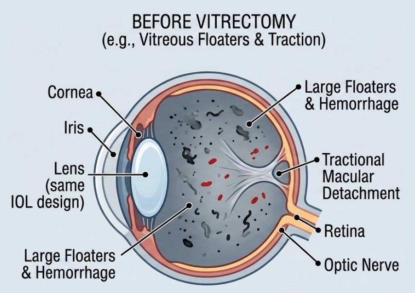
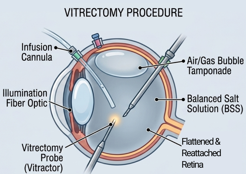
Surgery aims to reattach the retina and preserve vision. Whether the macula, the central part of the retina, was affected before treatment has a particular bearing on what is realistic, and we will discuss this for your situation.
These two procedures both involve peeling a very thin layer from the surface of the retina. They are described in turn below.
A macular hole is repaired with a vitrectomy, combined with peeling of a very thin layer on the retinal surface called the internal limiting membrane. A gas bubble then supports the hole as it closes.
Face-down positioning is often required after this surgery, helping the gas bubble press gently on the macula. The period can range from a few days to several weeks depending on the technique used, and we give you detailed guidance so you can plan for it at home.
Surgery can close the macular hole and improve central vision for many patients, though the degree of improvement varies from person to person.
An epiretinal membrane is a thin sheet of scar tissue that forms on the surface of the retina and can distort central vision. It is treated by careful peeling of the membrane, and in most cases a gas bubble is not needed.
Vision usually improves gradually as the retina settles, though some distortion may remain afterwards. We will discuss the likely range of outcomes with you before you decide to proceed.
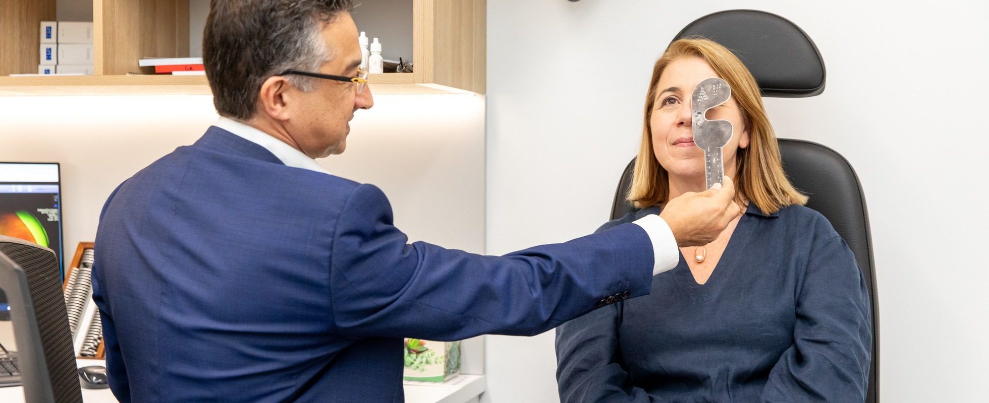
Recovery varies depending on which procedure you have had and whether a gas bubble was used. Your vision will be blurred at first and will clear gradually as the eye settles.
Some practical points to be aware of:
Please contact us or your referring doctor promptly if you notice sudden pain, a sudden increase in floaters, or any loss of vision after surgery. These changes warrant prompt assessment.
Every retinal operation carries some risk, and your surgeon will talk through these honestly with you before you decide to proceed. Knowing what can happen helps you give informed consent and recognise any warning signs early.
Possible risks include:
Most patients heal without serious problems, but no surgeon can guarantee a particular result.
We will explain the likelihood of these risks for your individual eye, weighed against the risk of leaving the condition untreated. This is part of the careful, individual assessment we give every patient.
Retinal surgery at our practice is provided by our retina team, who are specialist ophthalmologists with a subspecialty interest in retinal disease.
Before surgery, your eye is assessed in detail and your anaesthesia options are discussed with you. We work closely with your optometrist or GP throughout your care.
To arrange care, please contact us or read what to expect on our new patients page.
Most retinal surgery is performed under local or general anaesthesia, so you should not feel pain during the procedure. The eye may feel a little gritty or sore as it settles afterwards.
Recovery depends on the procedure. The eye heals over a few weeks, while improvement in vision, particularly after macular hole or epiretinal membrane surgery, can continue over several months.
Face-down positioning is most often needed after macular hole repair, as described in the macular hole section above. We give you clear instructions so you can prepare at home.
A gas bubble expands as air pressure drops, which can raise the pressure inside the eye to a harmful level. You must not fly until the bubble has cleared, and your surgeon will confirm when air travel is safe.
Retinal surgery is performed by our retina team, specialist ophthalmologists with a subspecialty interest in retinal and vitreoretinal surgery. You can also read about related care on our intravitreal injections page, where both injections and surgery may be relevant.
For further patient information, the Macular Disease Foundation Australia provides resources on retinal conditions and surgery.
The pages below cover the conditions these procedures treat, the injection treatments sometimes used alongside surgery, and the symptoms that often bring a retinal problem to light.
Retinal detachment, vein occlusion, macular hole, epiretinal membrane and uveitis, and how each one is assessed.
TreatmentInjections given into the eye for some retinal conditions, used on their own or alongside surgery.
Eye conditionWhat floaters and flashes mean, and the warning signs that need urgent assessment before a retina detaches.
Retinal surgery is a significant decision, and we encourage you to ask questions at every step. Ask your optometrist or GP for a referral, then get in touch to arrange your assessment. If your vision has changed suddenly, please say so when you call.
Retinal surgery at Eastwood Eye Surgery is provided by specialist ophthalmologists with AHPRA specialist registration in Ophthalmology and a subspecialty interest in retinal and vitreoretinal surgery. This information is general in nature and is not a substitute for personalised medical advice. Please speak with your ophthalmologist about your individual circumstances.