Eye condition
Retinal conditions are diseases that affect the retina, the light-sensitive layer at the back of your eye that makes sight possible. This page explains the medical and surgical retinal conditions we diagnose, monitor, and treat at Eastwood Eye Surgery, from gradual age-related changes to acute emergencies.
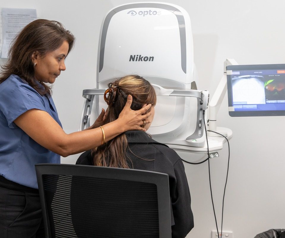
Retinal detachment: an eye emergency where the retina separates from the wall of the eye
Retinal vein occlusion: a blocked drainage vein that causes swelling and vision change
Macular hole: a small break in the central retina that blurs detailed vision
Epiretinal membrane: scar-like tissue that wrinkles the retina and distorts vision
Uveitis: inflammation inside the eye that can be isolated or linked to wider health conditions
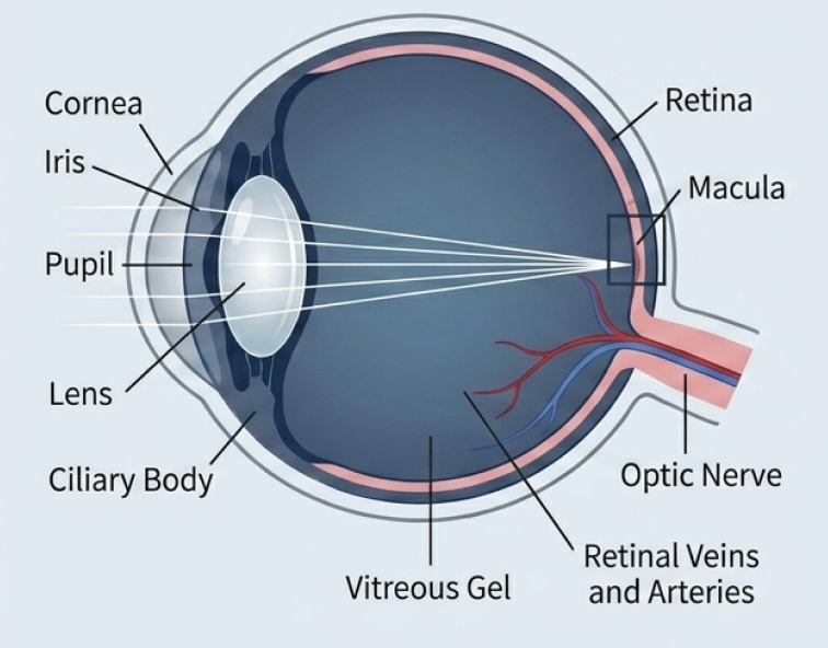
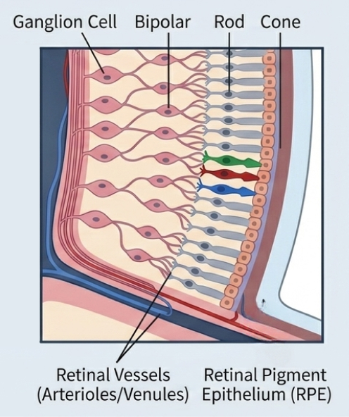
Retinal care at Eastwood Eye Surgery covers the full range of medical and surgical retinal disease. At every visit, we aim to:
The retina is the thin, light-sensitive layer lining the inside back wall of your eye. It captures the images you see and sends them to your brain through the optic nerve. The central part of the retina, the macula, is responsible for your sharp, detailed vision.
When the retina is damaged, inflamed, or pulled out of place, your vision can change quickly, which is why several of the conditions below need timely assessment.
Our retinal care is led by our specialist ophthalmologists with subspecialty training in retinal disease, including Dr Dov Hersh in medical retina and Dr Mark Gorbatov in medical and surgical retina.
You will need a referral from your GP or optometrist to see one of our specialists.
Some related conditions are covered in depth elsewhere on our site.
Retinal detachment is an eye emergency. If you experience sudden new floaters, flashes of light, or a curtain or shadow across part of your vision, seek urgent ophthalmology assessment immediately. Do not wait for a routine appointment.
Retinal detachment occurs when the retina separates from the supporting tissue beneath it, cutting off the oxygen and nutrients that retinal cells need to work.
Without prompt treatment, retinal detachment can cause lasting vision loss in the affected eye. When it is caught early, the retina can often be treated before lasting damage occurs.
The warning signs to watch for are:
These symptoms do not always mean the retina has detached. They can be the first sign of a retinal tear that may lead to detachment, so prompt assessment matters.
There are three main types of retinal detachment.
Some people are at higher risk. Risk factors include short-sightedness (high myopia), previous eye surgery, a family history of retinal detachment, and significant eye injury.
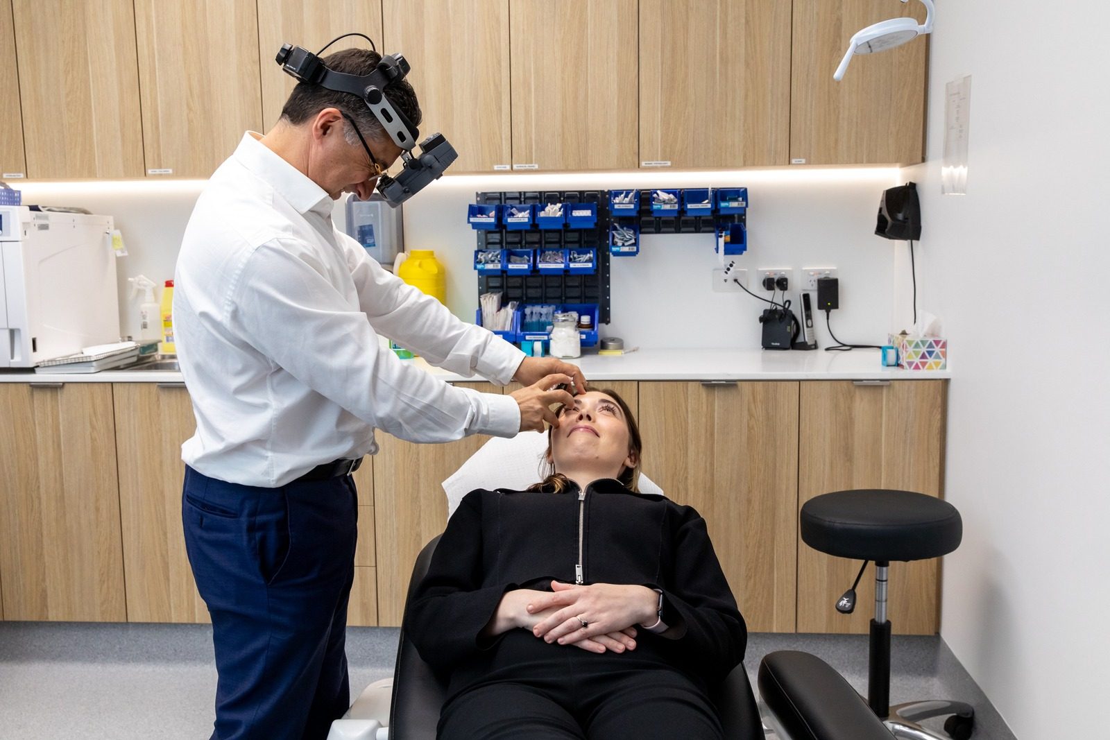
When you are seen urgently for these symptoms, your specialist will examine the back of your eye in detail.
This usually involves dilating the pupil with eye drops and using an instrument called an indirect ophthalmoscope to view the retina. If a clear view is not possible, for example because of bleeding inside the eye, a B-scan ultrasound may be used instead.
If you notice these symptoms, seek assessment urgently, ideally the same day, through your optometrist, GP, or our rooms during opening hours. Outside our hours, attend your nearest hospital emergency department.
Treatment depends on whether there is a tear alone or a full detachment.
Surgery aims to reattach the retina and preserve vision. Outcomes depend on the extent of the detachment and, in particular, on whether the macula was affected before treatment.
You can read more about these procedures on our retinal surgery page. If you have floaters or flashes but no curtain across your vision, our floaters and flashes page explains what to do.
Retinal vein occlusion is a blockage in one of the veins that drains blood away from the retina. When a vein becomes blocked, blood and fluid can leak into the retina, causing swelling and vision changes.
The blockage may affect the main central vein (central retinal vein occlusion) or one of the smaller branch veins (branch retinal vein occlusion).

The usual symptom is a sudden blurring or distortion of vision, most often in one eye. Some people notice the change on waking.
Vein occlusion is more common in people with high blood pressure, diabetes, or glaucoma. Part of the assessment therefore involves looking at your general health, not only your eye.
Treatment depends on how the occlusion is affecting your vision. Many cases are monitored closely with regular review and retinal imaging.
Where the blockage causes swelling at the macula, your specialist may recommend intravitreal anti-VEGF therapy, which involves injecting a medicine into the eye to reduce that swelling. You can read more on our intravitreal injections page.
Treatment aims to reduce macular swelling and stabilise vision for many patients, and managing the underlying health factors remains important.
A macular hole is a small break that forms in the macula. Because the macula provides your detailed vision, a hole there tends to blur or distort the middle of your sight while peripheral vision usually stays clear.
Macular holes most often develop with age. As we get older, the vitreous gel inside the eye shrinks and can pull on the macula, and this traction can open a hole over time.
A macular hole is not caused by anything you have done, and it is not related to macular degeneration, although the two are sometimes confused because both affect central vision.
The main treatment for a macular hole is a procedure called vitrectomy. This removes the vitreous gel and relieves the traction on the macula so the hole can close. You can read about this on our retinal surgery page.
Surgery aims to close the hole and improve vision for many patients. Outcomes depend on the size of the hole and how long it has been present, so your surgeon will discuss what is realistic in your case.
An epiretinal membrane, sometimes called a macular pucker, is a thin layer of scar-like tissue that grows across the surface of the macula. As this tissue contracts, it can wrinkle the underlying retina and pull it slightly out of shape.
Common symptoms are blurred or distorted central vision, with the sense that straight lines such as door frames or text appear wavy or bent.
The condition usually develops gradually, and many people notice it in one eye more than the other. It becomes more common with age and can follow other eye conditions.
Not every membrane needs treatment. If the membrane is mild and your vision is not significantly affected, your specialist may simply monitor it over time.
Where the membrane is distorting your vision enough to interfere with daily life, your surgeon may recommend vitrectomy with membrane peeling. This procedure removes the membrane from the surface of the retina, and more detail is available on our retinal surgery page.
Surgery aims to reduce distortion and stabilise or improve vision for many patients, and your surgeon will explain the likely benefit for your eye.
Uveitis is inflammation of the uvea, the middle layer of the eye that includes the iris, the ciliary body, and the choroid. It can affect one or both eyes and ranges from mild to severe.
Because inflammation inside the eye can damage delicate structures, uveitis is taken seriously.
Symptoms can include a red eye, eye pain, sensitivity to light, blurred vision, and floaters. A sudden, painful, red eye with light sensitivity warrants prompt review.
Uveitis sometimes occurs on its own. It can also be linked to wider health conditions such as ankylosing spondylitis, sarcoidosis, or inflammatory bowel disease.
Treatment depends on the type of uveitis, how severe it is, and what is driving it. Options range from anti-inflammatory eye drops to systemic anti-inflammatory medication for more extensive or recurrent disease.
The goals are to settle the inflammation, relieve symptoms, and protect your vision. We often work alongside your rheumatologist, GP, or physician to investigate any underlying cause and coordinate your care.
Common questions about retinal conditions are answered below. Your specialist can give you guidance specific to your eyes at consultation.
A retinal tear or detachment is the main emergency. The warning signs are set out above, under Acting Quickly on Retinal Detachment, and any of them calls for same-day assessment. If it is outside our opening hours, follow that same guidance and go straight to your nearest hospital emergency department.
Yes, you will need a referral from your GP or optometrist, as explained above. Your referrer can also help arrange urgent assessment if your symptoms have come on suddenly.
Often, yes. Many conditions are monitored with regular review and retinal imaging, and some, such as swelling from a vein occlusion, may be treated with injections into the eye. Surgery is reserved for conditions such as a macular hole, an epiretinal membrane affecting your vision, or a retinal detachment.
Not always. Floaters and flashes are common and often harmless, but a sudden increase can be the first sign of a retinal tear. Because it is not possible to tell the difference without an examination, any sudden change should be assessed promptly. Our floaters and flashes page explains what to do.
This depends on the condition and how quickly it is treated. Many retinal conditions can be stabilised, and vision preserved, when they are assessed and managed in good time, which is why sudden changes should not be left. Your specialist will explain what is realistic for your individual situation.
Sudden vision changes should be assessed promptly. If you are worried about a sudden change in your vision, contact us urgently so we can arrange the right assessment.
If you are a new patient, our new patient information page explains what to bring and what to expect. You can also reach our rooms through our contact page.
Our retinal care is provided by our specialist ophthalmologists. For independent patient education, Macular Disease Foundation Australia (opens in a new tab) provides reliable information.
The pages below cover the procedures used to repair the retinal conditions on this page, the injections used to treat macular swelling, and the symptom most often mistaken for a detachment.
The vitrectomy, scleral buckle and retinopexy procedures used to repair a detachment, close a macular hole or peel an epiretinal membrane.
TreatmentThe anti-VEGF injections used to reduce macular swelling after a retinal vein occlusion, and what the appointment involves.
Eye conditionWhat new floaters and flashes of light mean, and how to tell an ordinary change from an early warning sign of retinal detachment.
Further patient information:
RANZCO Eye Conditions (opens in a new tab) AAO Torn or Detached Retina (opens in a new tab)New floaters, flashes of light, or a curtain across your vision need urgent assessment. Ask your optometrist or GP for a referral, then get in touch with our rooms.
This information is general in nature and is not a substitute for personalised medical advice. Please speak with your ophthalmologist about your individual circumstances.